Description of the Movement:
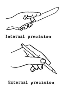
The dexterity of the hand is generally defined under two categories; power grips and precision grips. Precision unlike power grips are used with the intention of increased control as opposed to increased strength. Precision grips is then sub categorised into two groups (Figure 1);
- Internal: The shaft of the tool passes underneath the thumb; internal to the hand.
- External: The length of the tool passes over the thumb; external to the hand.
 Figure 1: Types of Precision Grips (Kanowski & Marras 1998)
The pinch grip is a form of precision grip where by an object is pinched between the palmar surface of the fingers and the opposing thumb. The pinch grip is also then categorised into five groups (Loudon, Swift & Bell 2008);
- Two-point tip pinch: Tip to tip grip of the fingers most commonly understood form of the pinch grip utilised when picking a pinch of salt for instance.
- Three-point tip pinch: The tip of the thumb met with the tips of digits 2 and 3 such as when holding a pen.
- Two-point pad pinch: The thumb and the pad of the index finger utilised to maneuver objects such as a light bulb
- Three-point pad pinch: The thumb opposes digits 2 and 3 with pads of the the distal phalanges such as during the opening (unscrewing) of a bottle cap.
- The-lateral pinch: A thin object held between the lateral surface of the index finger and the thumb such as when using a key.
Muscles Involved in the Two-Point Tip Pinch Grip:
During this movement the thumb is opposed and flexed while the fingers are flexed at the MCP, PIP and the DIP. In this grip compression is mainly produced by the extrinsic muscles of the hand (flexor digitorum superficialis, flexor digitorum profundus and palmaris longus) but also assisted by the metacarpophalangeal-joint flexion force of the interossei, flexor pollicis brevis combined with the adducting force of the adductor pollicis. Phalangeal rotation is adjusted by interossei and by the lumbricals. The opponens also provides assist through rotational positioning of the first metacarpal (Long et al. 1970).
 Figure 2: Anatomical position of Intrinsic muscles of the hand; provide fine motor control of the fingers primarily in the pinch grip through distal and proximal phalangeal flexion and adduction of the 1st digit through the use of thenar muscles (OpenStax College 1999).
Innervation:
Median Nerve:
The median nerve takes precedence in innervations of fine precision and pinch functions of the hand. It originates from the lateral and medial cords of the brachial plexus (C5-T1). During the pinch movement, the median nerve supplies digits 2 and 3 extrinsic flexion to the thumb; the motor branch supplying flexor digitorum superficialis while the anterior interosseus branch innervates the flexor pollicis longus and flexor digitorum profundus muscles.
The median nerve also supplies the thenar eminence. As the median nerve passes through the carpal tunnel, the recurrent motor branch innervates the thenar muscles (abductor pollicis brevis, opponens pollicis, and superficial head of flexor pollicis brevis). It also innervates the index and long finger lumbrical muscles. Sensory digital branches provide sensation to the thumb, index, long, and radial side of the ring finger.
Ulnar Nerve:
The ulnar nerve mainly takes part during power grips however it does supply the medial half of the flexor digitorum profundus (digits 4-5) through its motor branches, as well as provides sensation to the medial 1 and a half fingers of the hand. The ulnar nerve also takes part in the pinch grip by providing innervations to all interossei and 2 ulnar lumbricals; adductor pollicis and the deep head of flexor pollicis brevis (Wilhemi & Gest 2013).
Clinical Aspects:
If an individual is unable to pinch their index finger and thumb, a pathological indication of damage to the anterior interosseous nerve between the two heads of the pronator muscle is concluded. This is known as anterior interosseous nerve syndrome (AINS). AINS can be caused by compression of the nerve between the heads of the pronator teres muscle. The anterior interosseous nerve can also become prone to entrapment in select individuals with abnormalities such as an atypical head of the flexor pollicis longus muscle; found in approximately 52% of the population (Jones & Gallard 2014).
Try it yourself!
Do the ‘pinch test’ by forming an ‘OK’ sign with your thumb and index finger, being able to do this indicates your anterior interosseous nerve is functioning normally!
References:
Jones, J & Gallard, F 2014, RadioPaedia.org, accessed 25 April 2016, <http://radiopaedia.org/articles/anterior-interosseous-nerve-syndrome-1>.
Karowski, W & Marrras, WS 1998, Types of Precision Grips, digital image, The Occupational Ergonomics Handbook, CRC Press, London.
Long, C, Conrad, PW, Hall, EA & Furler SL 1970, ‘Instrinic-Extrinic Muscle Control of the Hand in Power Grip and Precision Handling’, The Journal of Bone & Joint Surgery, vol. 52, no. 5, pp. 853-867.
Loudon, JK, Swift, M & Bell, S 2008, The Clinical Orthopedic Assessment Guide, 2nd edn, Human Kinetics, United States.
OpenStax College 1999, Intrinsic Muscles of the Hand, digital image, Rice University, accessed 25 April 2016, <https://cnx.org/contents/FPtK1zmh@6.15:fEI3C8Ot@5/Preface>.
Whihelmi, BJ & Gest, TR 2013, Medscape, accessed 25 April 2016, <http://emedicine.medscape.com/article/1285060-overview#a6>.